GREAT WHITE SHARK DISSECTION
(Leigh Marine Laboratories)
accompanied by
MR images and renderings by the Digital Fish Library!
Leigh Marine Laboratories of Leigh, New Zealand had the rare opportunity to dissect an immature great white shark, Carcharodon carcharias. Weighing at 630 pounds and 9.8 feet in length, this female white shark drowned when it was caught in a gill net, and the fisherman who retrieved it generously donated the carcass to LML. Join Digital Fish Library researcher Kara Yopak and her colleagues at LML while they study one of the ocean’s most fascinating creatures.
*** DFL images from a younger white shark specimen
|
Storage
Where do you store a 630-pound great white specimen? Not in your average Coleman. After retrieving the specimen from the fisherman’s freezer shed, the big fish is transported and stored (many thanks to Halls Refrigerated Transport Ltd). The shark needs to be kept frozen to prevent the tissue from degrading. As you may have noticed, the management had a little fun with their 10-foot-long practical joke.
|
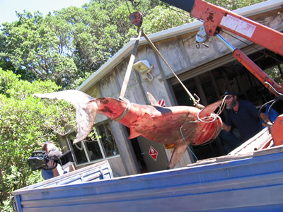
Transport
Before the dissection the shark is transported (via crane and a flatbed) to a shed at the LML where it can thaw. In these pictures you can really get a sense of scale. Imagine being in the water with a live one! (Remember, this shark isn’t even fully mature). After given some time to thaw, the shark is then hosed off and prepared for filming and dissection.
|
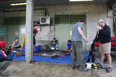
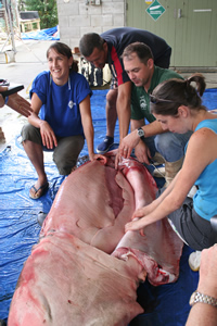
Preparation
Since an intact great white shark dissection is such a rare opportunity, National Geographic, as well as a host of other photographers, joins the scientists to document the event.
|
Parasites
Parasitic copepods are removed for further study. These animals attach themselves to areas of low velocity on the shark’s body (behind the pectoral fins, on the ventral side of the tail, etc). They are collected and will be sent to another laboratory for identification.
|
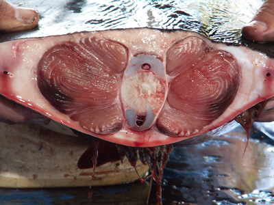
Red Muscle in Tail
This is where the white shark’s speed comes from. This cross section of the tail reveals two enormous bands of red muscle. These muscles power the tail from side-to-side, while the caudal keel slices through the water.
|
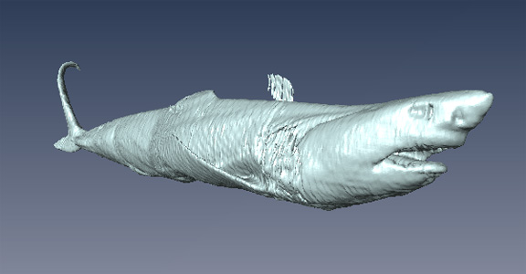
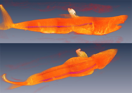
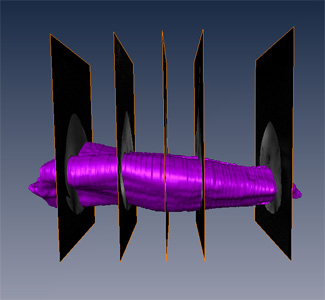
Red Muscle Along Length of Body (segmented MRI)
MRI allows us to view the two bands of red muscle in their entirety. Notice they run the length of the shark's body, from gills to tail.
|
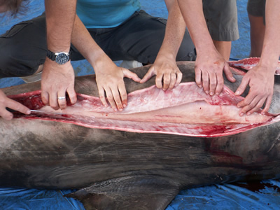
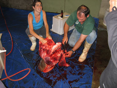
Gut Cut
All hands on deck as the dissection begins.
|
Liver
Cutting open the body reveals an enormous liver. In the absence of a swim bladder, sharks use their fatty liver for buoyancy. The liver also plays a major role in metabolism.
|
Liver (MRI)
2-dimensional MRI slices are stacked together to create a 3-dimensional rendering of the great white's huge liver.
|
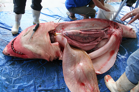
Internal Organs
Once the massive liver is heaved to the side, the stomach is revealed. Exploration of the stomach contents should prove to be interesting. White sharks do not have a discriminating palate. They eat a wide range of fishes and invertebrates as well as larger marine animals like porpoises, dolphins, seals, sea lions and even turtles and sea birds. Car tires and cuckoo clocks are among the items purportedly found in the bellies of these sharks – although these accounts have not been verified.
|
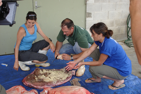
Stomach Contents
The stomach is opened up and the gut contents are examined. Maybe you can get an idea of the smell by studying the scientist’s faces. Kara Yopak describes the odor as “unlike anything you have ever smelled or will ever smell.”
|
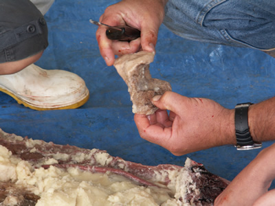
Last Meal
Clinton Duffy (Marine Conservation Unit, Department of Conservation, New Zealand) retrieves a whale vertebrae from amongst the partially-digested sludge. This will be sent to another laboratory for identification. Kara and Clinton speculate that this great white had been scavenging on a floating whale carcass. “Half-digested, rotting whale carcass is something I’m not too keen to smell again any time soon,” adds Yopak.
|
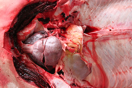
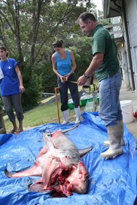
Heart
The stomach now properly disposed of, the scientists go on to examine the rest of the body. The pink organ shown here is the heart. Unlike humans, who have four-chambered hearts, white sharks and other elasmobranchs only have two.
|
Heart and Liver (MRI)
3-dimensional renderings of the heart (red) and the liver (purple). The liver makes up most of the shark's body weight.
|
Brain
After twelve consecutive hours of work, Kara gets to have a go at the brain. After taking initial measurements, the brain is carefully preserved and stored. Later, the five major brain areas (telencephalon, diencephalon, mesencephalon, cerebellum, and medulla) will be examined and compared to other shark species. Great white sharks have relatively enlarged mesencephalons and highly foliated cerebellums, similar to other sharks that primarily live in the open ocean.**
**White sharks were initially thought to be restricted to coastal waters, although recent research demonstrates that they can navigate huge distances through the open ocean.
|
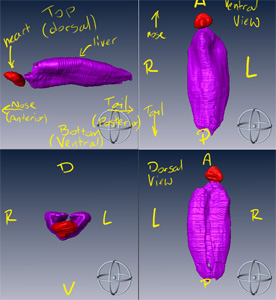
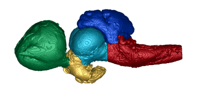
Brain (MRI)
Dorsal view of the brain of the great white shark. The forebrain, or
telencephalon, is shown in green and projects to the olfactory bulbs.
Further components of the brain are the diencephalon (yellow),
mesencephalon (cyan), cerebellum (blue), and medulla (magenta). The
relative size and complexity of these structures varies greatly between
species. The brain of the great white has a relatively large mesencephalon
(comprising 17% of the brain) and a highly foliated cerebellum (Yopak et
al., 2007).
|
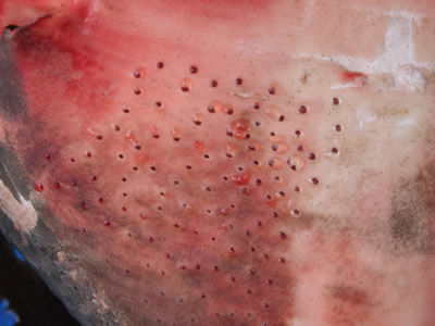
Pores of the Ampullae of Lorenzini
Notice these tiny jelly-filled pores on the snout and head. These pores are called ampullae of Lorenzini and are used to detect electro-magnetic fields. During muscle contraction, all animals produce a weak electrical field (think about going to the doctor for an EKG -- an electrocardiogram). These pores allow sharks to sense those weak electrical signals and detect fishes or other prey animals.
|
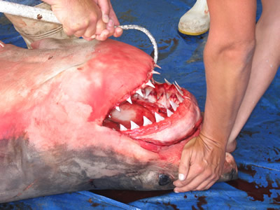
Jaws
White sharks deliver a powerful bite. Unlike humans, their jaws are not fused to the skull. When a white shark bites, it swings its jaws open, spits out its gums and bludgeons its prey with incredible force.
|
Great White Bite
An illustration of the jaw movements of the white shark (Tricas & McCosker 1984).
- The shark is at rest
- The snout is lifted and the lower jaw depresses
- The jaw protrudes forward, exposing the teeth and gums. Simultaneously the lower jaw moves up
- The snout drops, hammering the unlucky prey animal
|
Toothy Grin
Also notice the teeth. Remember that the jaws are unattached to the skull. Similarly, the teeth are unattached to the jaw. This allows for forward and backward rotation and retraction. These sharks have rows upon rows of pearly whites behind the main set. Any that are lost or broken off are easily replaceable. A close-up reveals the serrated edges, ideal for tearing flesh.
|
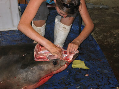
Cutting Out the Jaws
The fisherman who donated this shark was thoughtful enough to leave the jaws in place for scientific study, to be returned afterwards. When the dissection is over, the researchers remove the jaws to return to the fisherman as promised.
|
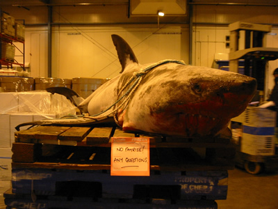
Another Specimen
Usually when marine laboratories receive great white specimens, they look more like this one. The jaws of the great white are valued at $10,000 to $20,000. This smaller specimen has already been picked over by the time it arrives at the laboratory.
|
Conservation
Great white sharks face considerable pressure from fishing and poaching. The monetary value of shark parts (the jaws, the fins), plus the status involved with catching such a notorious predator are among the many reasons these animals are currently listed as a threatened species on the IUCN Red List.
|